
Understanding Vascular Malformations
Vascular malformations, although relatively rare, can significantly impact an individual’s quality of life. These anomalies in blood vessels can present a plethora of challenges, from aesthetic concerns to serious health complications.
Understanding the different types of vascular malformations, their causes, symptoms, and treatment options is crucial for both patients and healthcare professionals alike.
In this comprehensive guide, we will delve into the intricacies of vascular malformations, shedding light on their various facets and how they can be effectively managed.
Types of Vascular Malformations
Vascular malformations encompass a spectrum of abnormalities affecting blood vessels. They are broadly categorized into five main types:
Venous Malformations: These malformations primarily involve veins and are characterized by dilated, tortuous vessels. They can occur anywhere in the body but are commonly found in the head and neck region. As the blood flow within them is slow, they are also called as low flow vascular malformations.
Arteriovenous Malformations (AVMs): AVMs are abnormal connections between arteries and veins, bypassing the capillary system. This results in high-pressure blood flow within the affected area, leading to various symptoms and complications.
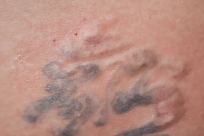
Capillary Telangiectasia: Capillary telangiectasia refers to small, dilated blood vessels near the surface of the skin or mucous membranes. While they may not cause significant health problems, they can be aesthetically displeasing and may bleed if injured.
Cavernous Malformations: Cavernous malformations are clusters of abnormally enlarged blood vessels that resemble a raspberry. They can occur in the brain and spinal cord, potentially leading to neurological symptoms such as seizures and weakness.
Lymphatic Malformations: Lymphatic malformations, also known as lymphangiomas, arise from abnormal development of the lymphatic system. They can manifest as cystic masses containing lymphatic fluid and may occur anywhere in the body, although they are commonly found in the head and neck region.
Causes and Risk Factors of Vascular Malformations
The exact cause of vascular malformations is often unknown, although they are believed to result from errors in embryonic development. Genetic factors may also play a role, as some malformations have been linked to specific gene mutations.
Additionally, trauma or injury to blood vessels during childbirth or surgery may contribute to the development of vascular anomalies.
Certain risk factors may increase the likelihood of developing vascular malformations.
These include a family history of vascular anomalies, genetic syndromes such as Klippel-Trenaunay syndrome and hereditary hemorrhagic telangiectasia (HHT), and exposure to certain environmental factors.
Symptoms and Diagnosis of Vascular Malformations
The symptoms of vascular malformations can vary widely depending on factors such as the type, location, and size of the anomaly. For example, individuals with venous malformations may experience pain, swelling, or discoloration of the affected area, particularly if the malformation is located in the extremities.
Also read – Effective Ways to Reduce Leg Swelling
In contrast, those with arteriovenous malformations (AVMs) may present with more severe symptoms such as pulsatile mass, warmth, or even neurological deficits if the AVM is located in the brain or spinal cord.
Diagnosing vascular malformations requires a comprehensive approach that often involves multiple healthcare professionals, including radiologists, vascular surgeons, and interventional radiologists.
The diagnostic process typically begins with a thorough medical history and physical examination, during which the healthcare provider may palpate the affected area and assess for signs of swelling, discoloration, or abnormal pulsations.
Imaging studies are crucial for confirming the presence of a vascular malformation and determining its characteristics. Ultrasound is often used as an initial imaging modality due to its non-invasive nature and ability to evaluate blood flow and vessel morphology.
However, more advanced imaging techniques such as magnetic resonance imaging (MRI) and computed tomography (CT) may be necessary to provide detailed anatomical information and assess the extent of the malformation.
In some cases, a biopsy may be performed to obtain tissue samples for histological examination, particularly if there is uncertainty about the nature of the lesion or if malignancy is suspected.
However, biopsy carries certain risks, such as bleeding or infection, and is typically reserved for cases where it is deemed necessary for diagnosis and treatment planning.
Treatment Options for Vascular Malformations
Once a vascular malformation has been diagnosed, the next step is to determine the most appropriate treatment approach. Treatment goals may vary depending on factors such as the patient’s symptoms, the location and size of the malformation, and the potential risks and benefits of each intervention.
Sclerotherapy
Sclerotherapy is a commonly used treatment modality for venous and lymphatic malformations, involving the injection of a sclerosing agent directly into the abnormal blood vessels. This causes irritation and inflammation of the vessel wall, leading to fibrosis and eventual shrinkage of the malformation. There are different sclerosing agents like Polidocanol, Sodium Tetradecyl Sulfate, Bleomycin and absolute alcohol which can be chosen according to the extent and location of vascular malformation.
Sclerotherapy is typically performed as an outpatient procedure and may require multiple sessions to achieve optimal results. However, while using absolute alcohol, which is painful, there is the need for hospitalisation and administration of anesthesia.
Embolization
Embolization is another minimally invasive treatment option that is particularly effective for arteriovenous malformations (AVMs). During embolization, a catheter is guided into the abnormal blood vessels supplying the malformation, and embolic agents such as Glue, Absolute alcohol, Squid or Onyx, coils or particles are deployed to block off the vessels and reduce blood flow.
Embolization can help alleviate symptoms such as bleeding, pain, or swelling associated with AVMs and may be performed as a standalone treatment or in conjunction with other modalities such as surgery or radiation therapy.
Surgical Resection
In cases where the vascular malformation is causing significant symptoms or complications, surgical intervention may be necessary to remove the lesion. Surgical resection may be performed using traditional open techniques or depending on the location and extent of the malformation.
Prevention and Management of Vascular Malformations
While it may not be possible to prevent vascular malformations entirely, there are steps that individuals can take to minimize their risk and manage their condition effectively.
Regular follow-up with a healthcare provider is essential for monitoring the progression of the malformation and identifying any new symptoms or complications early on.
In addition to medical management, individuals with vascular malformations may benefit from supportive therapies such as physical therapy, occupational therapy, or psychological counseling to address functional limitations, pain management, and psychosocial issues associated with their condition.
Also read – How to Manage Pain and Swelling
This multidisciplinary approach ensures that patients receive comprehensive care tailored to their individual needs and improves their overall quality of life.
FAQs About Vascular Malformations
An example of a vascular malformation is a venous malformation, which involves abnormally dilated and tortuous veins. These malformations can occur anywhere in the body but are commonly found in the head and neck region. Some birthmarks are also examples of vascular malformation.
Vascular malformations are developmental anomalies of vascular channels and will keep increasing with age and do not show any evidence of regression. They can also increase in size due to hormonal changes like puberty or pregnancy.
In contrast, hemangiomas are tumors with increase of vascular tissues and are typically found in infants, but they have rapid growth initially, followed by spontaneous regression. So more than 90% of hemangiomas will regress by the age of 7 years.
Whether a vascular malformation is life-threatening depends on various factors such as its location, size, and associated complications. While some vascular malformations may cause significant symptoms and complications that can be life-threatening if left untreated, others may be relatively benign and pose minimal risk to health.
Vascular malformations typically do not go away on their own. They are considered congenital anomalies resulting from errors in embryonic development and tend to persist throughout life unless treated. However, some vascular malformations may remain stable or undergo minimal changes over time, while others may progress and cause worsening symptoms or complications.
Whether a vascular malformation causes pain varies depending on factors such as its location, size, and involvement of surrounding tissues. Some individuals with vascular malformations may experience pain or discomfort, particularly if the malformation compresses nearby structures or affects nerve endings. However, not all vascular malformations cause pain, and some individuals may be asymptomatic or only experience mild symptoms.
Whether a vascular malformation causes pain varies depending on factors such as its location, size, and involvement of surrounding tissues. Some individuals with vascular malformations may experience pain or discomfort, particularly if the malformation compresses nearby structures or affects nerve endings. However, not all vascular malformations cause pain, and some individuals may be asymptomatic or only experience mild symptoms.
Conclusion
In summary, vascular malformations encompass a wide spectrum of anomalies within blood vessels, each presenting distinct challenges. From venous malformations to lymphatic malformations, understanding these variations and their symptoms is pivotal for accurate diagnosis and effective treatment.
With advancements in medical procedures such as sclerotherapy, embolization, and surgery, tailored care is available to improve patients’ quality of life and reduce complications. Managing vascular malformations necessitates a collaborative approach involving specialists in radiology, vascular surgery, and interventional radiology.
For those experiencing symptoms or diagnosed with vascular malformations, seeking guidance from a seasoned professional is paramount. Dr. Sumit Kapadia offers personalized consultations and treatment plans tailored to individual needs.
With his expertise and compassionate care, Dr. Kapadia aims to guide patients toward optimal health and well-being. Schedule a consultation with Dr. Kapadia today to embark on the journey toward effective management of vascular malformations.

MBBS, MS, MRCS, DNB-Fellow
Dr. Sumit Kapadia
Dr. Sumit Kapadia / MR KAPADIA SUMIT a gold-medalist from Baroda Medical College, obtained his general surgical training and senior residency from SSG Hospital, Vadodara.
MBBS, MS, MRCS, DNB-Fellow
Dr. Sumit Kapadia
Dr. Sumit Kapadia / MR KAPADIA SUMIT a gold-medalist from Baroda Medical College, obtained his general surgical training and senior residency from SSG Hospital, Vadodara.
